The Science Behind the Goose Bump

The skin is the largest organ in the human body. Most people don’t realize the critical role skin plays in the survival of multi-cellular organisms. The skin is air tight and provides a mechanical and defensive barrier between the outside environment and the body’s internal structures. It is what holds us together, gives us shape, if you will, and this multifaceted structure is perhaps the less appreciated out of all the body’s organs. The skin contains blood vessels, nerves, sweat glands, specialized glands to ward off pathogens, immune cells, sense receptors, hair, and muscle. And that’s not even the half of it. Within the skin’s three distinctly different layers, its components are specifically  arranged to produce a dynamic organ with the capabilities to repair itself. It is for these reasons, and many more, that makes the skin one of the most interesting organs of the body.

Many of the skin’s functions developed out of our need to survive. For example, the malanocyte is a specialized skin cell that helps the body ward off ultraviolet radiation by darkening our skin.

skinAnother evolutionary adaptation is the goose bump, that prickly raising of the skin that occurs when one is cold or senses danger. What exactly is the goose bump, and why is it important? Let’s take a look at a  simplified cross-section of human skin. As mentioned, mammalian skin has three layers. The epidermis, the dermis and the hypodermis. Each hair is contained in a sheath of epidermal cells called a hair follicle. This follicle is associated with sebaceous glands, capillaries, and a microscopic smooth muscle bundle called the arrector pili. This tiny muscle bundle attaches the hair follicle to the underside of the epidermis.

Let’s now look at the mechanics of the goose bump formation. Ordinarily, the hair emerges from the skin at an angle. When the arrector pili muscle fibers contract, this action pulls the lower half of the hair follicle toward the epidermis, resulting in the hair standing more erect. The result of this creates a “bumping” of the skin surrounding the shaft of hair above the skin. You now have a goose bump.

So, that’s the mechanics, now let’s take a look at the cause of a goose bump. Goose bumps are the effect of activation of the sympathetic branch of the autonomic nervous system. As mentioned above, the skin is innervated by nerves. Nerve cells respond to stimuli in the body and produce neurotransmitters, small molecules that bind to receptors on a cell surface.  The arrector pili muscle cells contain alpha 11) adrenergic receptors which bind to the neurotransmitter norepinephrine. The release of norepinephrine from the neuron follows the “fight-or-flight” response. When a person senses danger, surprise, or the body is under certain stress, the neuron receives a message and responds by releasing nor-epi. Norepinephrine then binds to the cell surface, and series of events occur within the cell that causes the arrector pili to contract.

Why do we have goose bumps? The answers are quite simple. Our ancestors had more hair than we did. To withstand the frigid temperatures the arrector pili was stimulated, causing the hairs to stand on end and trapping warmer air closer to the skin. It provided our ancestors with the thermal layer they needed. Erect hair also shows dominance. This is seen today when the hairs of a dog stand on end. Perhaps long ago our ancestors displayed the same sort of aura to their rivals. Imagine 150,000 years ago if you were face to face with your enemy who had hair sticking out like a pekingese dog. My guess would be you would do more than laugh. In a more practical sense, when someone becomes frightened or threatened, the “fight-or-flight” response kicks in, and a host of reactions take place. Heart rate increases, digestive functions decreases, skeletal muscles contract, eyes dilate, blood flow increase, hair stands on end. This reaction helps us fight, or allows us to get out of the way fast. It most likely is a reason for the success of Homo sapiens as a species.

The average adult has about 5 million hair follicles. That’s an awful lot of goose bumps.

The Virus: The Accidental Organism

As science continues to progress, our understanding of life is evolving, thanks in part to the advances in molecular biology. Scientists can now take a closer look at organisms than ever before. Not only has the Linnaean classification system (Kingdom, Phylum, Class, Order, Family, Genus, Species) gone out of favovirus 2
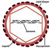
r with many scientists due to the molecular unraveling of evolutionary histories of the species, but also the idea of what constitutes a living organism has been put on trial.

Living organisms grow, reproduce, and carry on biological processes within their cells. From the more complex organisms, such as mammals, to the one celled paramecium, life processes are conducted throughout the growing species’ lifetime. Viruses represent that unique biological entity bordering on the threshold of life. It has long been established that viruses exhibit none of the characteristics mentioned above, and the molecular study of them has begged the question are viruses alive?

In order to address this question, let’s first take a look at the structure of a virus. All viruses contain two things: a genome and a capsid (the protein coat that surrounds the genome). The genome can be DNA or RNA, single-stranded or double-stranded. The capsid varies in shape from rod-shaped to a more complex icosahedron. Although a few viruses contain unique enzymes, most viruses contain nothing else. Some viruses are surrounded by a lipid bilayer containing glycoproteins. These glycoproteins bind to specific receptors on the surface of the host cell, making the virus cell specific. Viruses that do not contain a lipid bilayer may have glycoproteins attached to the capsid. The binding of the virus via its glycoprotein to the host cell’s receptor initiates entry of the virus into the cell.

Viruses are obligate intracellular parasites, that is they can only survive within the cell. They have no cytoplasm, no nucleus, no mitochondria or other cellular organelles. They are incapable of making their own energy or manufacturing proteins. The virus relies on the host cell’s proteins/enzymes, ribosomes, and energy to replicate its DNA/RNA and to manufacture its capsid proteins, which are translated from its own genome. Viruses are assembled within the host cell from preformed components. They require no nutrition. Because of this, the virus exhibits no change during its life time. From the moment it forms and buds from the host cell, to the time of its “uncoating” in the next cell it infects, it is exactly the same.

One may wonder where these submicroscopic entities come from in the first place? There is speculation that they arose from life forms that have lost cellular functions. Another theory, and the one I support, is that viruses evolved as a result of the macromolecule (DNA or RNA)  escaping the confines of the cell. Viruses range in size from 20nm – several thousand nm. Their genome ranges from ~6kb (about 10 genes) to ~1.2 Mbp as seen in the Mimivirus (possibly >900 genes). This is small compared to the genome of living organisms (E. coli >5,000 genes, humans ~21,000 genes). The Mimivirus is unique in that many scientists consider it a bridge between the nonliving virus and living organisms.

The question whether the virus is living or not will be under debate for some time. There is still much to learn about the submicroscopic organisms, and until we unravel the mysteries, these small, seemingly non-living particles will continue to infect and destroy their hosts. Many of the nucleic acids found in viruses have the propensity to integrate into the hosts’ genomes and are responsible for the onset of many cancers, a topic for another day. Make no mistakes, the virus is the perfect vehicle for transporting unwanted DNA or, as in the case of the HIV virus, RNA throughout the body.

virus binding

To Barbecue, or not to Barbecue.

photo-45

Summer is finally upon us. After weeks of rain and unpredictable weather, we are now able to enjoy those favorite summer activities. Swimming, biking, kayaking, …and oh, let’s not forget the barbecue. What a more perfect way to spend with family and friends than to be outdoors cooking on the grill. But make no mistakes, there are health issues to consider before you fire-up that grill.

It has been well established that cooking meat on the grill is dangerous for your health. Deadly, as a matter of fact. A high consumption of meat that has been cooked over an open flame or on grills that use hydrocarbons does indeed contribute to the onset of cancer. Studies have shown an increase in colon cancers in persons who eat food cooked in this way. Why is this, and what is the culprit?

Before I answer those questions, it’s important to understand the nature of cancer. Cancer is not a single disease. There is an enormous number of genes whose alteration will initiate cancer in a particular cell type. Processes within a normal cell are highly regulated. The highly ordered sequence of events in the life of a cell is called the cell cycle. Within this cycle, there are signals that tell cells when to grow, divide and die. If these signals are altered, the cell cycle becomes unregulated and cancer ensues.

Cancer starts with a mutation in DNA. The induction of the mutation varies depending on the carcinogen (radiation, chemical, biological or endogenous). Among the most common include DNA insertions or deletions, breaks, dimers, and the addition of adducts to a DNA strand. DNA replication proteins become confused by these changes and replicate the DNA strand erroneously. As a result, the protein coded by the mutated gene does not function normally, or the protein becomes silent or absent within the cell.  Sometimes the cell recognizes this abnormality and the DNA is either repaired or the cell dies in the process known as apoptosis. Other times the cell escapes this fate and continues to divide and live longer than it ordinarily would because the protein(s) that once kept it in check are no longer functioning normally. The cell also loses its ability to adhere to other cells and migrates through the body (a process called metastasis).

The cancer causing chemical found in grilled foods is called benzo(a)pyrene (BaP). BaP is a polycyclic aromatic hydrocarbonBaP that forms as a result of incomplete combustion of organic molecules. Simply put, the burning of any organic substance, such as cigarettes, wood, gasoline, meat, may create BaP. When meat is cooked on a grill, molecules normally found in meat can be transformed into BaP. BaP can also be formed from the fuel used by the grill and be carried to the meat with the smoke. As a result, BaP covers grilled meat. Then you eat it. A marshmallow roasted over an open flame also contains BaP.

BaP is not as harmful to humans as its metabolites. BaP enters the blood stream, and once it enters a cell it is converted to other compounds by specific enzymes. The resulting metabolites bind to DNA, forming large adducts that confuse the proteins responsible for DNA replication. Most of the time these adducts are removed through DNA repair mechanisms, but not always. If enough of the adducts remain, the protein coded by the mutated DNA strands does not function normally, or it is simply not transcribed at all. There will then be a cascade of events that triggers the onset of carcinogenesis.

The effects of BaP is cumulative. That is, the more BaP metabolites that bind to DNA, the more likely your chances of getting cancer. It is simply a matter of numbers: it’s more efficient for a DNA repair protein to remove a single adduct from DNA than it is for it to remove a dozen. What this means is that if you absolutely love char grilled food, than eat it. But do so in moderation.

Life Depends on the Stomata

A few months ago I wrote a post entitled “How Insects Breathe.” The purpose of that post was to illustrate just how varied the respiratory processes are for animals. And it gets stranger. Gas exchange in an earthworm occurs across their skin as these organisms do not have respiratory organs. Fish, as we all know, have gills, which extract oxygen from the water as it flows opposite in direction to its blood. Regardless if the species is an insect, a human, an earthworm or a fish, the end result is the same. Oxygen enters the body,  then diffuses into the cells, where, along with sugar, goes through a series of pathways to produce CO2 , water and energy through a process called cellular respiration. Plants also carry on cellular respiration using the same pathways and processes. Let’s take a look at the chemical reaction for cellular respiration in plants and animals. (The energy produced is not listed here):

er4

But here’s the clincher. The starting material for respiration is glucose. Plants do not consume sugars, so they must be made by the plant before glycolysis, the Krebs cycle and oxidative phosphorylation occurs. And they do not “breathe” in oxygen, which is also needed for cellular respiration. The plant must produce its own oxygen.  The starting material for cellular respiration in plants is produced through the process of photosynthesis. Let’s take a look at the chemical reaction for photosynthesis (Again, minus the energy needed to kick off the reaction):

en2

Look familiar? It should because it is cellular respiration in reverse, but make no mistakes, other than the fact that the starting and end products of these two processes are reversed, there is nothing similar to them at all. One may speculate that the two processes could become a cycle where their products are recycled over and over again, and that would be the case if it weren’t for the fact that much of the oxygen that is produced in photosynthesis readily leaves the systems. And that is a good thing, otherwise the sugar made by the plant would never be stored and used for our consumption, but would instead be broken down and animals would not be able to survive.

So, how does oxygen leave the system? Or, more precisely, how does oxygen get out of the plant? That task takes place through tiny apertures called the stomata. Stomata are found on the stem, some flower parts, and leaves. In most deciduous plants, stomata are found primarily on the underside of the leaf. The number of stomata varies, and there may be as many as 40,000 per square centimeters on some leaves.

The aperture of the stoma is surround by two specialized cells called guard cells. The alteration in the shape of the guard cells widens to allow CO2 uptake into the plant, and O2 is released intst0ma 2
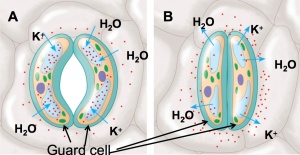
o the atmosphere. Let’s take a look at how this happens. The concept is simple. When water enters the guard cells, the cells swell and become turgid, and because they are attached at their ends, the space between them widens. Why does water enter the guard cells in the first place? Water diffuses from a low to a high concentration of solute. Potassium (K+ ) first enters the guard cell from neighboring cells, and as a result, water follows and the stomata open. This is shown at A on the drawing to the left. When K+ leaves the guard cells, water follows, the guard cells become flaccid and the stomata close. This is illustrated at B.

Along with the O2, water also exits the plant by way of the stomata. This process is called transpiration, and if left unregulated, the plant will wilt. The job of the plant is to maximize fixation of CO2 into sugars while minimizing water lose. During times of drought or high temperature, the roots produce abscisis acid (ABA), which travels through the plant and signals the guard cells to close. Problem solved. Almost. When droughts persist, the stomata remain closed, CO2 does not enter, the plant does not grow. Thus is the cause of failed crops during times of drought. Sunlight also effects the opening and closing of the guard cells. It would make sense that during daytime when the sun is brightest the stomata will open, because photosynthesis occurs in sunlight. The closing at night will minimize water lose within the plant.

The stomata are one of many fascinating structures found in a plant and can be seen if the underside of a leaf is careful pealed off and place under a microscope. They just might be the most important biological structure on the planet, because without them CO2 will not enter the plant and sugars, the molecule that keeps our cells working, will not be made. The existence of life on Earth may very well indeed be dependent upon the stomata.

The Origin of Mitochondria

We have all been taught that mitochondria are the powerhouse of the eukaryotic cell (cells containing membrane bound organelles such as the nucleus, golgi apparatus, mitochondria …). It is within this membrane bound organelle where most of the energy needed for our cells to survive is produced. What exactly is this energy, and how did mitochondria come about?

The energy, quite simply, is ATP (adenosine triphosphate). ATP is produced by a series of pathways involving many enzymes, cofactors, and of course glucose. With every glucose molecule that enters a cell, roughly 36-38 molecules of ATP are produced. Glucose begins its breakdown in the cytoplasm of the cell through the anaerobic pathway of glycolysis. The products of glycolysis are then transferred into the mitochondria where they are shuttled into the Krebs cycle and then on to oxidative phosphorylation. In respect to cellular respiration, the difference between the cytoplasm and the mitochondria besides the enzymes that are present, is the use of oxygen. Aerobic respiration, which consumes oxygen in the reaction, only occurs in the mitochondria. It is the addition of this extraordinary organelle that aided in the evolution of the eukaryotic cell and therefore the higher species.

mitochondria

It is believed that mitochondria arose through a process called endosymbiosis nearly 2.1 billion years ago. This theory suggests that a bacterium entered a cell, lived in a mutually beneficial relationship with its host and hence evolved into the mitochondria that we are familiar with today. The diagram at the left illustrates how this could have happened. In A, the cell membrane surrounds a bacterial cell and engulfs it. In B, we see that the bacteria now contains an outer membrane, compliments of the host cell. The original bacterial plasma membrane becomes highly folded and becomes the inner mitochondrial membrane that we see in C. This folded structure increases the surface area where oxidative phosphorylation takes place, therefore many of these reactions can occur simultaneously, optimizing the amount of ATP produced. Quite an efficient system for something that had once been a lowly bacterium.

The evidence for this theory is overwhelming. First, mitochondria are similar in size and shape to some strains of bacteria. They are between 0.5-1 μm in diameter and 1-10 μm long. The inner membrane of the mitochondria contain enzymes similar to those found in some bacterial cells. Mitochondria also contain their own genome. It is a circular molecule like that found in bacterial cells. mtDNA containing ~16 kilobases. Thirty-seven genes have been identified. Thirteen of these genes encode proteins used in cellular respiration that takes place within the mitochondria, and 24 code for RNA molecules involved in mitochondrial protein synthesis. There is a greater degree of homology between mitochondria RNA and the RNA found in bacteria than there is between mtRNA and the RNA found in the cytoplasm of eukaryotic cells. Mitochondria reproduce within the cell by way of fission, as do bacteria.

Mitochondria are inherited by the mother. They are found in the egg, and any mitochondria that might have been inside the sperm and entered the egg during conception are destroyed. Because of this, maternal linages can be easily traced.

The number of mitochondria within a cell type varies considerably depending on the energy needs of that cell. As you may imagine, muscle cells and brain cells contain thousands because their energy needs are greatest. Red blood cells do not contain any mitochondria, as their life is short and their energy requirements are produced in their progeny cells.

Without mitochondria, the organism would not be able to meet its energy needs and we might still be one cell organisms swimming in an oxygen deficient bog.

ENDORPHINS. Not the happy drug you’re looking for.

The human body has many mechanisms that signal that something is not right. For instance, a fever indicates the presence of a harmful pathogen, thirst is a natural response to dehydration, and feeling pain brings about an awareness that damage is occurring within the body. In turn, the body may release hormones or other molecules that help overcome the crisis.

We have known for some time that endorphins act as a natural analgesic, that is, it is a natural pain reliever.  They are considered endogenous opioids because they bind to the same cell surface receptor as do opium and morphine, and hence have similar effects. There are many myths surrounding the release of endorphins into the blood stream. Many people believe that endorphins are released when one is happy, during orgasms, even when someone smiles. The truth is, that there is NO scientific evidence supporting these claims. In fact, the level of endorphins in the blood stream has been shown to decrease during sex8.

Endorphins are released in the body in response to pain, or extraneous exercise. Simply put, they prevent someone from feeling the pain associated with tissue damage. β-endorphin is a 31-amino-acid-peptide that functions as a neurotransmitter, that is, its target cell is a neuron. β-endorphin is released from the anterior pituitary gland in response to a pain signal that has been sent to the hypothalamus.  It then enters the blood stream.

Let’s first look at the pain signal. There are pain receptors throughout the body, called nociceptors. When tissues are damaged, they release a variety of substances, such as histamine, potassium and arachidonic acid. These substances stimulate the nerves and cause the release of Substance-P, which activates the pain pathway and transmits the pain signal to the central nervous system. In this way, the brain not only recognizes pain, but it also commits its source to memory. It is this recognition that creates the reflex to move away from the source of the pain.

endorphin

The opiate receptor is located on the neurons that release Substance-P. When β-endorphin is release in response to pain, it will bind to this receptor and as a result Substance-P will not be released. Therefore, the pain pathway will not be initiated, or will be reduced. β-endorphin is quickly degraded by an enzyme, and the receptor will then be free to bind to its substrate again. If this degradation process did not take place, then the feeling of pain will be nullified and the hand that inadvertently sat on the hot stove will remain there.

Addiction to opioids such as morphine and opium is the result of the drugs remaining on the receptors for long periods of time. A person becomes numb after a while because the receptors become “down regulated,” that is, there are less of them on the surface of the cell. Quite simply, there are fewer receptors and it takes more of the drug to find them. It then takes more of the drug to reach the same level of the “pain free” high.

The physiological role of endorphins in the human body has not yet been fully elucidated. However, one thing is clear. Its role is not to induce happiness, but to remove the pain.

Crossing the Cell Membrane

When I first decided to write a post about membrane transport,  it seemed like an uncomplicated topic to tackle. But once I began the actual writing process, I realized that the subject was far too intricate to cover in a one page blog post. After all, cell membranes are responsible for maintaining ion concentrations between the cell and its environment. They provide a mechanical barrier that traps cellular constituents unique to each cell type. And they are selectively permeable and regulate what molecules enter the cell and what molecules leave the cell. The cell membrane is a complex, multi-functional structure that communicates with matrices inside and outside the cell as well as with other cells.  So for the sake of simplicity, I will focus on only a few types of transport processes and save the more complicated energy dependent multi-step transfers for another time.

phosp

Before talking about membrane transport,  let’s first take a look at the membrane’s basic structure shown at the left. A cell membrane is comprised of a phospholipid bilayer with the polar (hydrophilic, or “water-loving”) heads facing into the cell (intracellular) and outside of the cell (extracellular). The non-polar (hydrophobic, or “fat-loving”) ends of the phospholipid face inside the bilayer. This bilayer structure completely surrounds the cell and is filled with proteins, cholesterol and glycoproteins.  Many of the proteins transverse the membrane, some do not, and they are in constant communication with other proteins in the complex extracellular and intracellular matrices.  Membranes are fluid, that is they are in constant motion, and it takes about 1 second for a phospholipid to travel the length of a bacterial cell (about 2 μm).

Let’s take a look at the one-dimensional depiction shown below on the left. This over-simplified drawing illustrates the lipid bilayer with three proteins embedded into it, but keep in mind that this is hardly representative of what is actually there. The lipid bilayer acts as a cell membrane
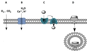
“gate-keep,” allowing only small, hydrophobic molecules such as O2 and CO2 to move through it unaided through the process of passive diffusion. This is illustrated in “A.” In this way, O2 is delivered quickly and efficiently into the cells when needed and its entry doesn’t depend on the presence of a protein, which might ultimately slow down the process because it takes a while for the proteins to make their way to the cell membrane. Despite their size, steroid hormones, such as testosterone and progesterone, can also pass through the cell membrane this way because they are fat-soluable and are not impeded by the membrane’s hydrophobic interior.

Ions and small polar molecules such as water require a transport protein to facilitate their movement through the membrane. The interior of the transport protein is charged, thereby attracting these molecules. This is illustrated at “B.” Each protein is unique in that it will only transport one type of molecule. For instance, the transport protein shuttling H+ across the membrane is different from that used for Ca++ .  Many of these proteins, such as  the aquaporins used to transport water molecules, are brought to the cell surface only when needed.

The drawing at “C” illustrates a transport protein that undergoes a conformational change upon the binding of its substrate. Similar to those shown at “B,” these proteins are specific for one particular substance, and many are brought to the surface only when needed.  As shown in the picture, the substrate binds to the binding site of the transport protein, and this interaction causes a “flip-flop,” or conformational change in the protein that results in the release the molecule into the cytoplasm. This release then causes the protein to resume its original structure. Glucose enters the cell by this means. There are several types of glucose transport proteins, and many of them are tissue specific. For example, the glucose transport proteins found in the brain are not dependent upon insulin. They are embedded in the cell membrane at all times to ensure the brain receives a constant supply of glucose regardless of the individual’s diabetic status.

The last mode of membrane crossing illustrated here is called receptor-mediated endocytosis, the process by which cells internalize large molecules that bind to cell surface receptors. This is illustrated  at “D.”  Once a molecule binds to the receptor on the cell surface, the membrane will invaginate, encasing the bound molecule within its own membrane. Enzymes take over from there, and the molecule is released into the cytoplasm and the membrane and receptor may then be recycled back into the cell membrane, thereby maintaining the size of the cell. Insulin, some vitamins and the HIV virus are examples of substances that gain entry into the cell by this method.

The survival of every organism is dependent upon this complex bilayer of their cells. One genetic mutation may prevent a particular protein from making its way to the cell membrane and result in serious consequences to the life of the organism. I am in constant awe at that premise, and once again it reveals that the mammalian species is truly remarkable.

Thanks for stopping by!

The Nature of DNA

Untitled

All living things grow and reproduce, and there must be a blueprint, directions if you will, that enables the organism to carry out these processes and maintain the integrity of the species. That blueprint, in most organisms, is deoxyribonucleic acid, better known as DNA. There are some viruses that do not have DNA, and instead use RNA as their blueprint. HIV is one of them. But many scientists argue that a virus is not a living organism, a topic to be discussed at a later date.

The structure of DNA was elucidated in 1953 by James Watson and Francis Crick. I had the honor of meeting Dr. Watson, the only survivor from that era, and I must say it was like meeting Mic Jagger. Watson stands as a symbol to perhaps the greatest achievement known to science, but make no mistakes, there were others involved in the discovery who never reaped the honors bestowed upon Watson and Crick. Rosalind Franklin deduced the structure of DNA prior to Watson and Crick through her X-ray diffraction photograph. Sadly, she passed away in 1958 at the age of 38 before the Nobel prize was awarded.

So what exactly is DNA? All proteins in the body are coded from DNA through a multistep process. DNA is a polymer, a molecule comprised of repeating similar parts. It is a double-stranded helix containing a series of the 5-carbon sugar deoxyribose, a nitrogenous base and a phosphate group. The phosphate group is what makes DNA acidic.

DNA 1

Let’s take a closer look at the double-stranded structure on the right. The backbone of DNA consists of alternating sugar and phosphate groups. The two strands run antiparellel. The sugar is chemically bonded to a nitrogenous base, which in turn forms hydrogen bonds with its complimentary base, forming complimentary base pairs. There are four different nitrogenous bases in DNA: Adenine (A), Thymine (T), Cytosine (C), and Guanine (G). A always pairs with T, and G always pairs with C. This molecule can replicate itself through a complex series of steps involving enzymes that are coded using its DNA. It’s the replication of this molecule that maintains the integrity of the dividing cell, and ultimately the species. If it were replicated erroneously, then the proteins that are coded from it will be altered, and they will not perform their job very well. As a result, genetic disorders may occur, as well as cancers.

DNA is separated into genes, the unit of hereditary information that codes for one particular protein. It is the sequence of nitrogenous bases, commonly known as the nucleotide sequence, that denotes what protein will be translated from that particular gene. Only one strand of the DNA codes for a protein, and that nucleotide sequence must remain constant. For example, a section of the nucleotide sequence that partially codes for a particular DNA repair protein is 5′ – GGC AAT CCT GTC CCC ATC – 3′. 5 Altering just one of these bases (creating a point mutation) may decrease the activity of this protein, or render it nonfunctional. That could be devastating, considering its job is to repair damaged DNA, or it could be beneficial in cases where you want to destroy the cell by way of DNA modulation, as in cancer chemotherapy. If just one point mutation in one particular gene could cause such drastic consequences, it is a wonder the human species is sill thriving. But there are many safeguards in place to circumvent the consequences caused by DNA damage, and that is what makes the cell so fascinating.

The human body is very complex and each cell contains 20,000 – 25,000 genes, and roughly 50 – 250 million base pairs. This is a lot of DNA, but fear not, DNA is efficiently packaged in the nucleus of most cell types (the exception being prokaryotic cells, which include bacteria).  Before cell replication, the genetic material condenses further to form chromosomes. Each chromosome is comprised of a single strand of DNA,  measuring on average 1.5 x 108 base pairs, that is tightly coiled and packaged along with proteins called histones. There are 23 pairs of chromosomes in human somatic cells (any cell except a sperm or egg). That makes 46 chromosomes per cell. Lined up end to end, the DNA in those chromosomes would extend approximately 2 meters in length. Not bad for a string of molecules.

histones

The understanding of DNA has come a long way from the days of Watson and Crick, but we still have miles to go until we fully unravel this marvelous structure.

How Insects Breathe

UntitledFor my first post, I would like to say something about insects. There are an estimated 10 million species of insects on Earth, and their success may be attributed to their simple, yet efficient, body structure. Their segmented exoskeletons, a topic we will discuss at a later date, provides armor that protects their bodies from trauma that would otherwise harm them. They have complex social structures and are fascinating creatures to watch.

Insects do not have lungs, but like other organisms within the Animalia kingdom, they require oxygen to carry on life processes.

Insects breathe through tiny pores on the sides of their abdomens. These pores, known as spiracles, are found on each segment of the abdomen and open and close in response to the concentration of oxygen within the insect’s tissues. They also close to prevent water loss within the insect. Just like humans, when an insect respires there is an exchange of gases, (O2 for CO2) and a loss of water.  When air passes through the spiracle, it will then travel through a trachea. These tracheae connect in a pipe-like network of air tubes  that extends the length of the insects body. They terminate in close association with an insect cell where the gas exchange will then take place between the membrane of the tracheoles and the cell membrane through the process of diffusion. This system of respiration is considered the simplest and most efficient respiratory system in animals. Besides the spiracles found on the abdomen, most insects also have three pairs of spiracles at the base of their legs.

Insects belong to the phylum Arthropoda. Most terrestrial arthropods (spiders,  ticks, centipedes) have this system of respiration. Aquatic arthropods, such as lobsters and crabs, breathe by a specialized internal gill system.

Please feel free to comment below, or if you wish to add anything, please do.